

Home > Research > Research Highlights > Biology > Even Cells Have a Social Infrastructure: Understanding Intracellular Logistics
date: 2018.09.10
Associate Professor: TAMURA Yasushi (Molecular Cell Biology)

▲Fig.1
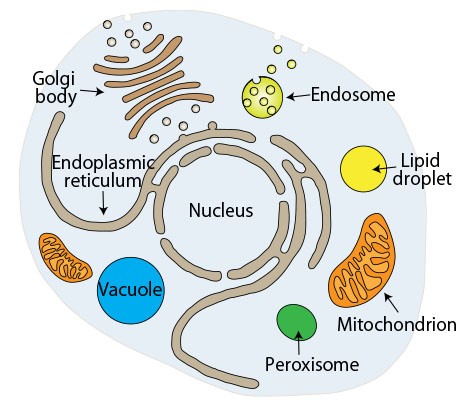
Structures called "organelles" develop inside the cells which compose a human body. Organelles are surrounded by lipid membrane(s). Main examples of organelles are the nucleus which stores DNA, the endoplasmic reticulum which performs protein synthesis, and mitochondria which produce the energy needed to live (Fig.1). Abnormalities in organelles as the "social infrastructure of cells" are known to cause a variety of human diseases.
"Intracellular logistics systems for transporting an appropriate amount of necessary substances to the right place at the right time" are essential to maintain the social infrastructure of cells. Since long ago, the intracellular transportation mechanism of proteins has been actively studied and many important points have been clarified. On the other hand, the intracellular transportation mechanism of lipids has not been investigated well because it is difficult to experimentally handle lipids due to the water insolubility. However, given that organelles composed of lipid membranes (Fig.2) play critical roles in the social infrastructure of cells, research on lipid logistics is essential to understanding cellular society.

▲Fig.2
We have discovered the proteins responsible for transporting lipids from the mitochondrial outer to inner membrane. We have also clarified that the proteins which directly bond mitochondria and the endoplasmic reticulum, conduct lipid transportation in the range between the two membranes.

▲Fig.3
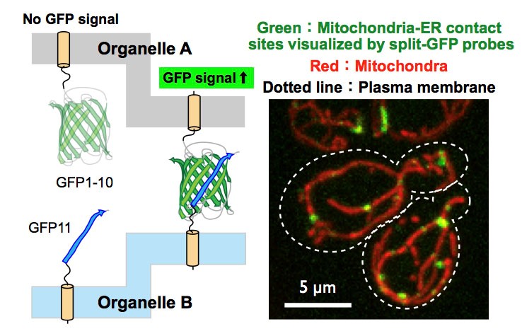
Until now, organelles were thought to exist independently. The discovery of how organelles form contact sites overturned this conventional wisdom and had a huge impact. Now, the question is whether other inter-organelle contact sites exist inside of cells. Also, assuming that such sites exist, what are the binding factors between organelles? In order to answer these questions, we must conduct experiments to evaluate (visualize) inter-organelle contact sites. As the result of trial-and-error, our research group succeeded in visualizing contact sites between different organelles by using a split-GFP system which consists of fluorescent proteins that have been divided in two (Fig.3). By utilizing this visualization technique, we hope to discover new inter-organelle binding factors and clarify new functions in inter-organelle contact sites.
Related Links